Intraoperative MRI
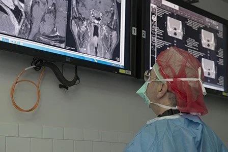
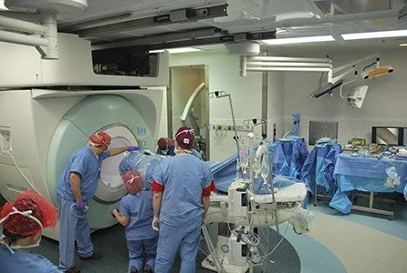
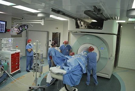
The Neurosurgical Service moved into the new Lunder operating rooms in 2012. At that time, a 3-Tesla state-of-the-art intraoperative MRI was incorporated into two of the operating rooms. This allows the surgeon to image patients under anesthesia during their operative procedures. It allows near- real time evaluation of the adequacy of resection during the procedure itself. This is especially valuable in cases like pituitary tumors, where the entire tumor cannot be easily visualized
Patient undergo transsphenoidal surgery in the usual fashion using the microscope or endoscope. At the conclusion of the resection, the MRI magnet, which is housed in a special shielded room, is brought into the operating room. High-quality 3-Tesla images are obtained under anesthesia. The images are reviewed by the surgeon and radiologist. If additional resectable tumor is seen, the surgeon can re-explore and remove the additional tumor. This technology allows us to obtain maximal tumor resection.